Share on Facebook
 Share on X
 Share by Email
 Share on Google Classroom
Medical ultrasound (synonyms: diagnostic ultrasound, sonography, ultrasonography) is the use of high frequency sound waves that are far above the range of human hearing to image the interior of the body. It is used widely in Canada in both hospital and out-patient settings to assist in the identification and/or treatment of many diseases, and to monitor pregnancy. Therapeutic ultrasound, the use of sound waves in physical therapy to provide deep heating to soft issues such as muscles and tendons, is a different process and is not the subject of this article.

History
The piezoelectric effect was discovered in 1880 in Paris by the brothers Jacques and Pierre Curie. They observed that certain crystalline materials, such as quartz, generate an electric charge when subjected to mechanical stress, or external force, thus converting mechanical energy to electric energy. The reverse effect also occurs. If an electric charge is applied to the crystalline material, it emits mechanical energy in the form of sound waves. The sinking of the Titanic in 1912 and the onset of the First World War in 1914 spurred efforts at the detection of hazardous icebergs and enemy submarines respectively. During those years Canadian-born investigator Reginald Fessenden developed an underwater acoustic ranging device capable of detecting an iceberg 3 km away. However, it did not use the piezoelectric effect and was not suitable for submarine detection. In 1918, the French physicist Paul Langevin used the piezoelectric effect to develop a successful submarine detection device that used a quartz crystal to both emit and receive high frequency sound signals. This led to the development of sonar (sound navigation and ranging) systems, which played an important role in combatting submarine warfare during the Second World War. By harnessing the piezoelectric effect in a device that both emitted and received sound waves, Langevin and his team laid the groundwork for the later development of medical ultrasound transducers.
During the late 1930s and into the 1940s, many investigators in different fields studied the ability of sound waves to interact with various body tissues as a means of diagnosis. These included the study of the motion of heart valves and the brain. A common problem facing many researchers was that the sophistication of their ideas exceeded that of the equipment available to them. American radiologist Douglass Howry headed a team that constructed a functional “home-built” ultrasound scanner in 1949. His pioneering work, along with that of Ian Donald, is considered a direct precursor of today’s scanners. Donald, an obstetrician in Glasgow, Scotland, and Tom Brown, an engineering colleague, developed a scanner suitable for the examination of pregnant women in the mid-1950s, leading to the recognition of Donald as the “father of obstetrical ultrasound.” Also in the 1950s, ultrasound was used to study tumours in the breast and the brain and to diagnose retinal detachment. The work of these and other pioneers began to generate interest in the medical community, which in turn spawned commercial interest in the manufacture and sale of ultrasound scanners. Concurrently, the development of digital electronics and powerful microprocessors led to the development of advanced instruments and a multi-billion-dollar global market.
Underlying Physics
Medical ultrasound is based upon the basic pulse-echo principle: a pulse of sound is emitted in a known direction, strikes a target, and returns to the emitting source, where it is registered. If the velocity of sound in the medium is known, the distance to the target can be calculated from the round-trip travel time. The returning echo must be registered before the next pulse is emitted. It is assumed that the echo travels along the same straight line as the emitted sound.
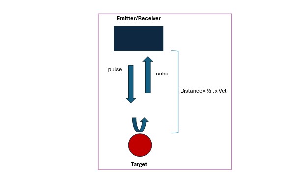
In order to conduct a medical ultrasound examination, an operator places an ultrasound transducer on the skin, applying a thin layer of gel to eliminate air pockets between the skin and the face of the transducer. The transducer’s piezoelectric elements emit short bursts, or pulses, of sound that travel through body tissue at an average velocity of 1,540 m/sec. The returning echoes are received by the transducer and analyzed by the computer in the scanner as to strength, direction and time of arrival from tissue boundaries. Typically, 1,000 to 10,000 sound pulses are emitted per second (the pulse repetition frequency), enabling the construction of a cinematic two-dimensional display of the region being examined in real time. For medical purposes, ultrasound frequencies between 2 and 15 million cycles per second (2–15 MHz) are used. Lower frequency sound waves penetrate farther into the body before undergoing loss of energy than those of higher frequency. For deeper organs, such as the liver or kidney, a 3–5 MHz transducer is usually required, whereas a 7.5 –10 MHz transducer is used for superficial structures such as the thyroid gland. The compromise is that the images produced by lower ultrasound frequencies are of lesser resolution than those of higher frequency, and do not show as much fine detail.
There is a complex interaction between ultrasound waves and body tissues that depends greatly upon the composition of the tissue. The degree to which a material transmits ultrasound is known as its “acoustic impedance” and is related to its density. When sound strikes a boundary between tissues of similar acoustic impedance, most of it passes readily across the boundary and a weak echo is generated; if there is a large acoustic impedance difference at the boundary, most of the sound will be reflected, creating a strong echo. Strong echoes are displayed as white on the ultrasound image, weaker echoes as shades of grey, and absence of echoes as black. This display is known as real-time, grey-scale, B-mode (brightness mode) imaging and is the mainstay of medical ultrasound.
When the ultrasound beam strikes a boundary at an oblique angle, it may be refracted, or bent, from its path, returning to the transducer at a delayed time and along a path that is different from the emitted axis. This “fools” the scanner into creating a false image, or artifact. Other physical processes can also create artifacts, all of which can potentially lead a poorly trained observer to a wrong diagnosis.
Uses of Ultrasound
Ultrasound is used widely because of its portability, safety record, relative low cost compared with CT and MRI scanning, and because it does not use ionizing radiation (X rays). The latter property makes it ideal for obstetrical imaging. The Society of Obstetricians and Gynaecologists of Canada recommends that all pregnant women have a “dating” ultrasound at 11–14 weeks after the last menstrual period and an “anatomic” ultrasound between 18 and 20 weeks. Lack of radiation also renders ultrasound very useful in the assessment of neonates and young children.
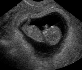
Ultrasound is used to examine the abdominal and pelvic organs; the breasts; the heart; blood vessels; and superficial structures such as the thyroid gland, salivary glands, scrotum, lymph nodes, and ligaments and tendons. It is employed to guide interventional procedures such as organ biopsies, drainage of abscesses and other fluid collections, the placement of catheters in blood vessels, and injections into joints. Small ultrasound transducers can be placed in the rectum, vagina, blood vessels, heart and digestive tract for very specific indications. As a general principle, ultrasound is of limited use in the assessment of bone and of gas-filled structures.
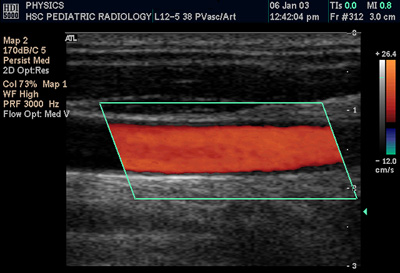
Doppler sonography assesses blood flow in the heart, arteries and veins. When an ultrasound beam of a known frequency strikes a moving target such as flowing blood, the returning echoes will undergo a frequency shift that enables the scanning device to determine the speed and direction of blood flow.
The Doppler frequency shift can be encoded in colour for ease of rapid, real-time assessment. The Doppler apparatus is often combined with B-mode imaging in what is called “duplex imaging.”
Cardiac ultrasound, or echocardiography, is used to assess heart function. “M-mode” (motion mode) ultrasound may be added to the standard B-mode imaging to evaluate the motion of rapidly moving structures such as the heart valves and chamber walls. Three-dimensional “(3-D) ultrasound” is the volume rendering of ultrasound data to add depth to the conventional 2-D image. Among other applications, it can be helpful in assessing the fetus for certain congenital anomalies.

Canadian Context
Ultrasonography is an essential component of medical imaging in Canada. Although many individuals have contributed to its development, the following physicians are recognized as pioneering practitioners, investigators and educators: Drs. Edward Lyons, Peter Cooperberg and Stephanie Wilson. Medical ultrasound has many strengths, but one limitation is its operator dependence. Inadequately trained individuals, both physicians and technologists (sonographers), can be a source of incorrect diagnoses. Most physicians who practise ultrasound are certified with the Royal College of Physicians and Surgeons of Canada and have taken advanced ultrasound training. They interpret and report upon the documented observations of the sonographers. Canadian sonographers have typically completed several years of classroom and practical study at an accredited educational institution. Sonography Canada is the national body that provides professional practice guidelines and promotes excellence for sonographers.